Spectra is an open source and safe way to experiment with biomedical imaging. It has reconstruction algorithms, a PCB and can re-create an image in real time.
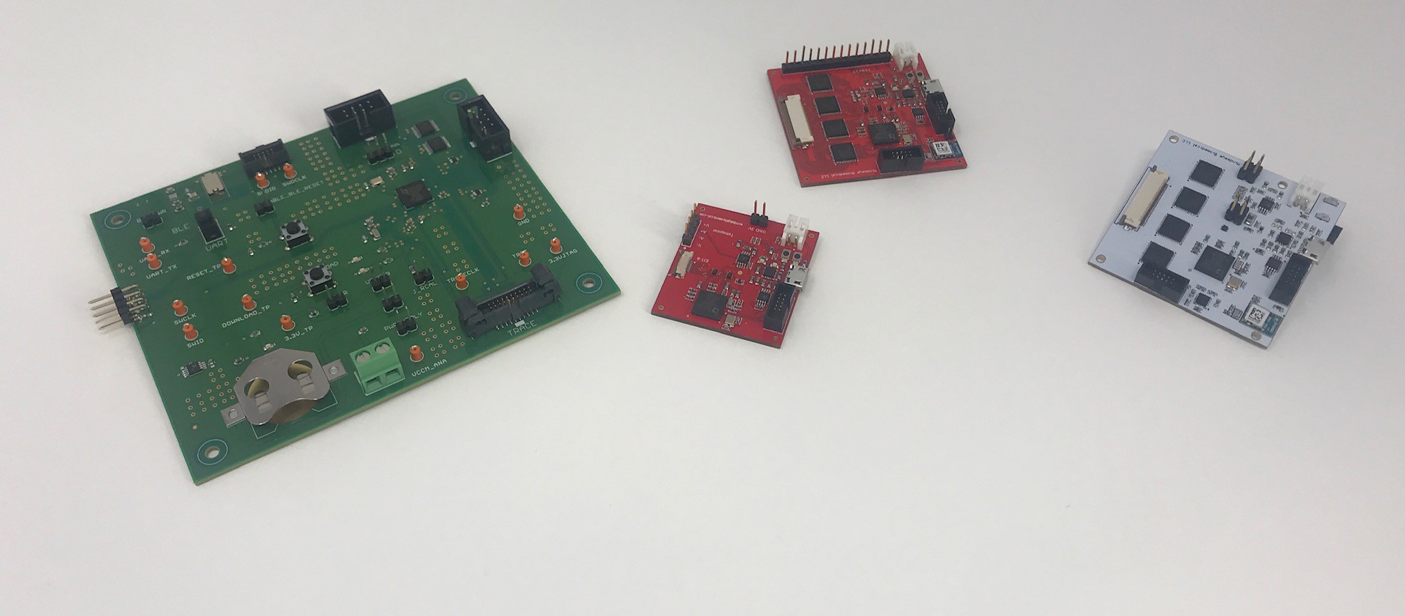
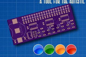
An AC current is sent through the conductive medium to be interrogated, and the impedance magnitude and phase is measured at the other electrode. This process is repeated around every combination of an array of electrodes, which gives us the base data to perform a tomographic reconstruction. The board is a tiny 2" square, with bluetooth, 16 bit resolution and 160kSPS sample rate. It does calibration based on a resistor on the board, which also adjusts for environmental temperature differences to remain accurate.
Below is a video of an image being reconstructed in real-time using only 8 electrodes. Notice the image moves as the shot glass rotates. Since then we've moved to a smaller PCB design with 32 electrodes!
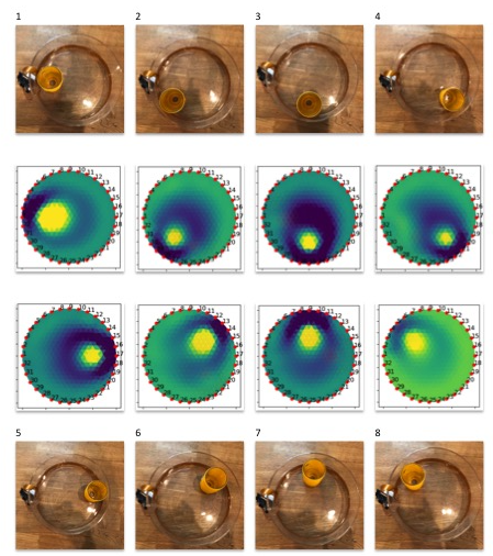
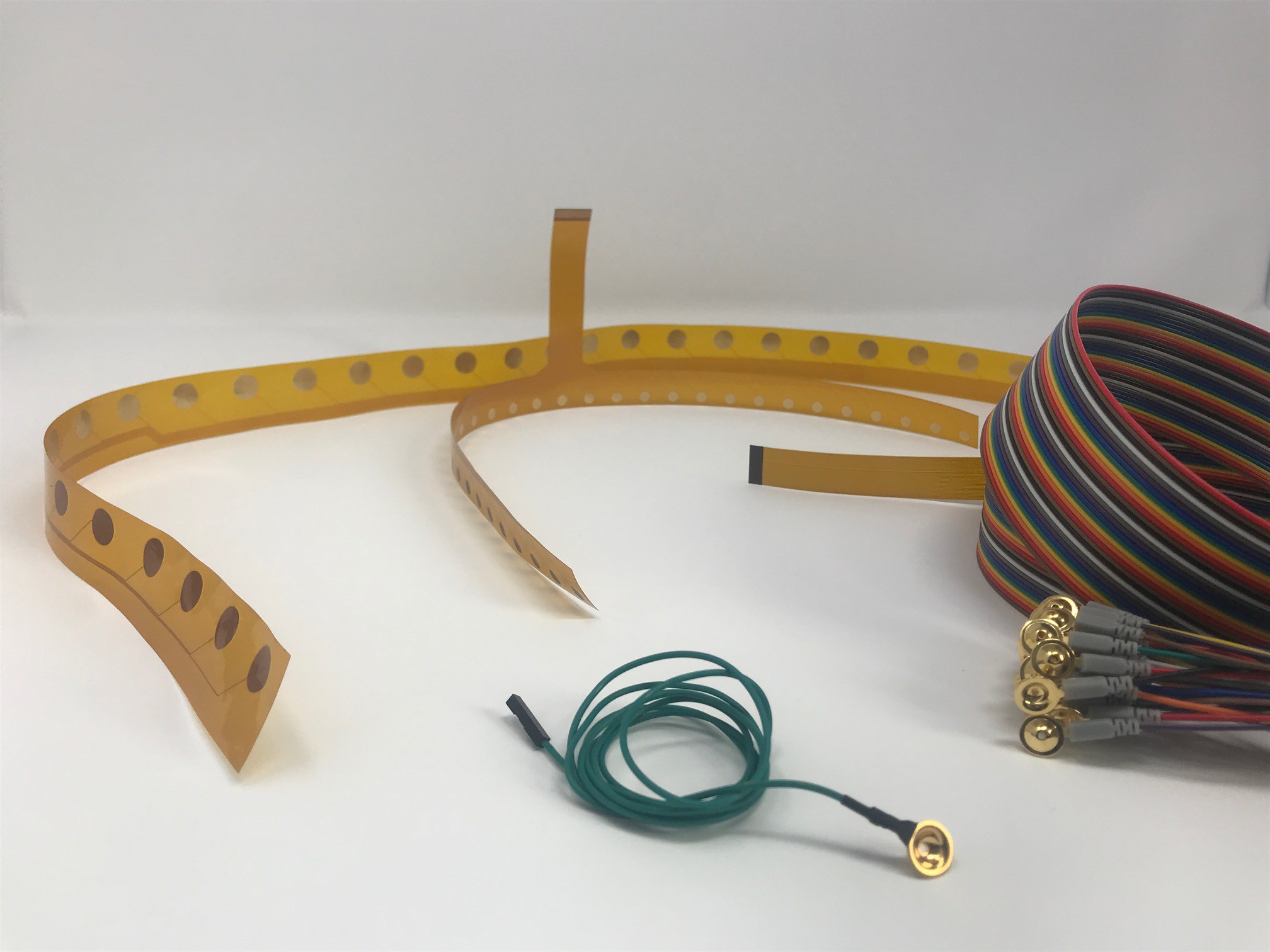
32 electrodes gets better spatial resolution than 8 electrodes, but is slower as it takes 896 combinations in total per image. You can easily switch reconstructions algorithms, edit and tune them in software and try again from Gauss Newton, Graz Consensus to good old fashioned Back Projection(the same algorithm as a CATSCAN).
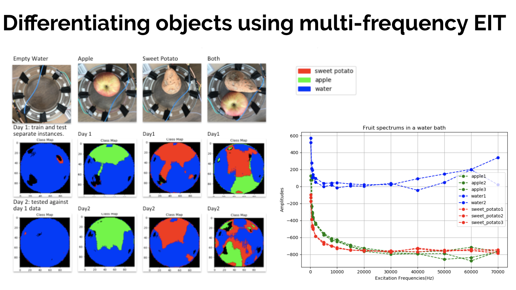
You can easily experiment with reconstruction algorithms or see if you can detect differences in materials such as fruit shown below. Each piece of fruit has it's own unique dielectric signature, based on the cell characteristics within it. You could also apply this to meat, bones, blood clots or tumors just as easily. Since this spectrum data is collected at every spatial location, it makes it harder to visualise and ideal for a multidimensional tool like machine learning. People are only just beginning to apply machine learning techniques here so it seems ripe for easy improvements.
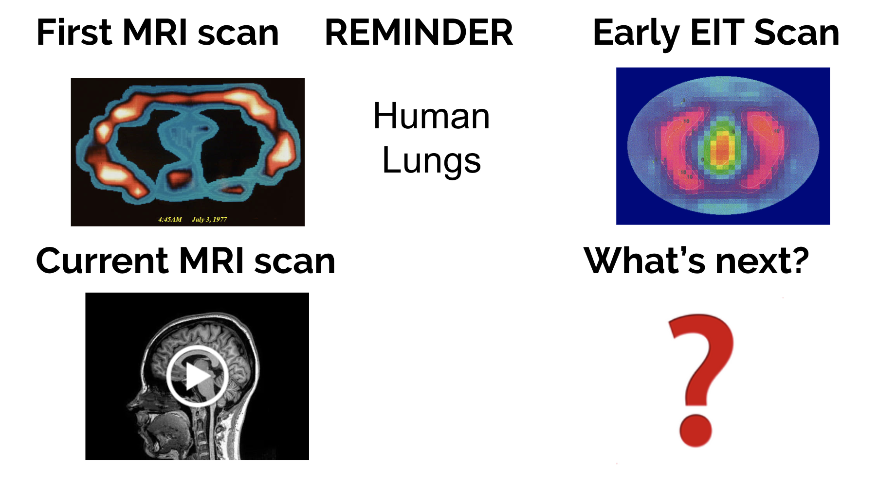
MRI started with some pretty rough images too with the original thorax picture shown below. MRI's still cost millions of dollars, and are never going to be so small an accessible that anyone can have one and play with it. Below we see the first ever MRI scan of the human thorax, with a current 3 Tesla scan below it to show how far the technology has progressed. EIT is in its early stages, so it will be interesting to see how it improves.
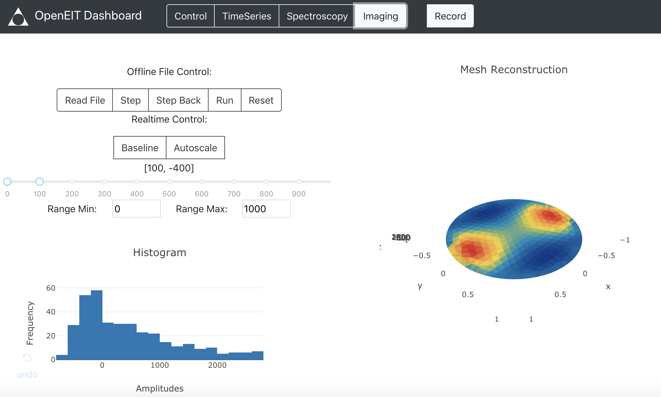
Here is a link to the project on github, that contains an easy to use dashboard with reconstruction algorithms that can be run in real-time or alternatively read in offline files. There are 3 different reconstruction algorithms currently available. The true strength of this technique I think is that you can get a dielectric spectrum at every pixel, meaning you can see differences in material properties really well.
The project from python tomographic reconstruction software to hardware and firmware is open source. Please refer to the readme to install it! Questions and collaborators welcome:
 jean
jean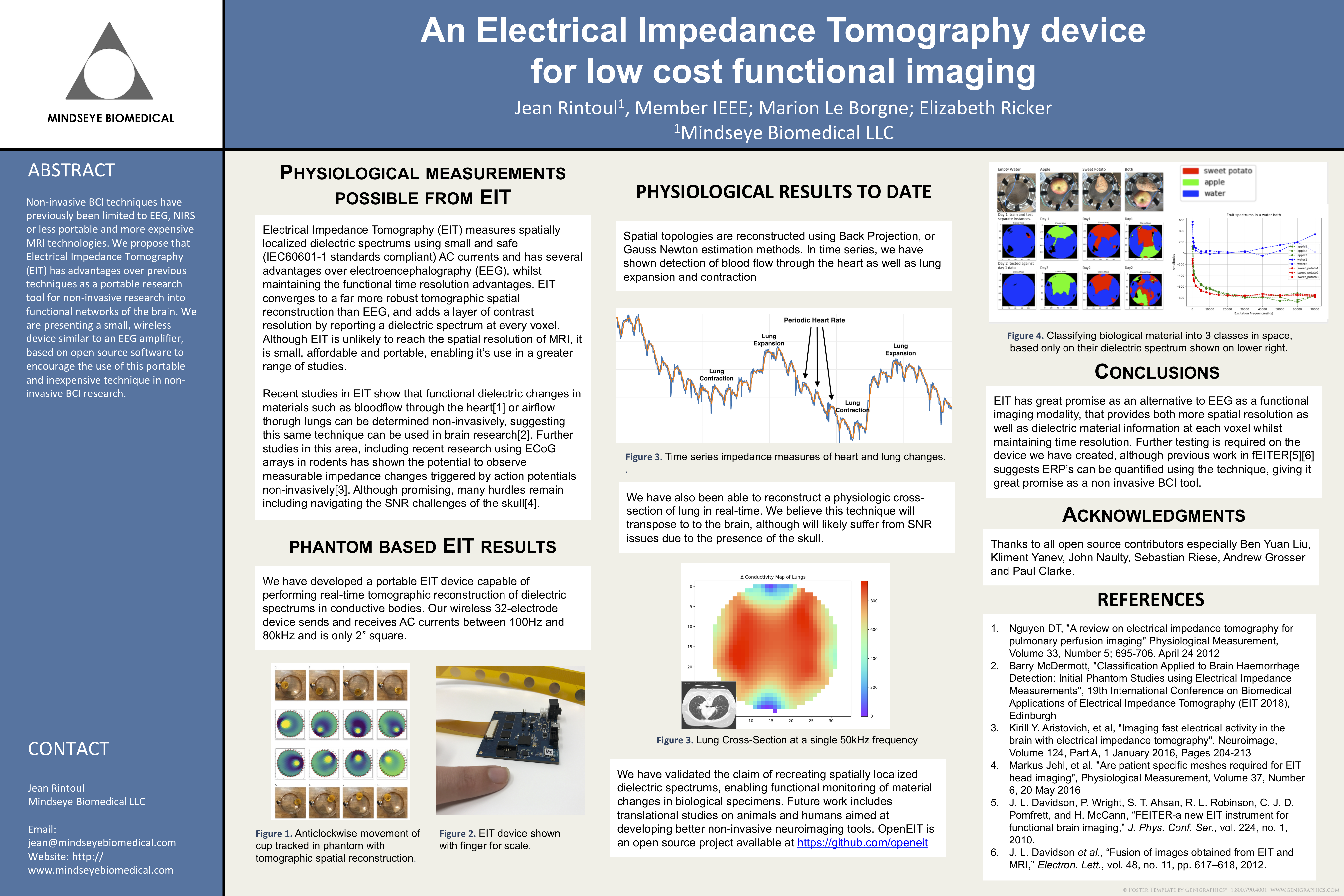




 Eric Wiiliam
 Dan Berard
 jens.andree
 James Pilgrim
Congratulations team Spectra! Enjoy those coffees while you whip the dev kits into existence!