Once you have the photos, the magic maths comes in!
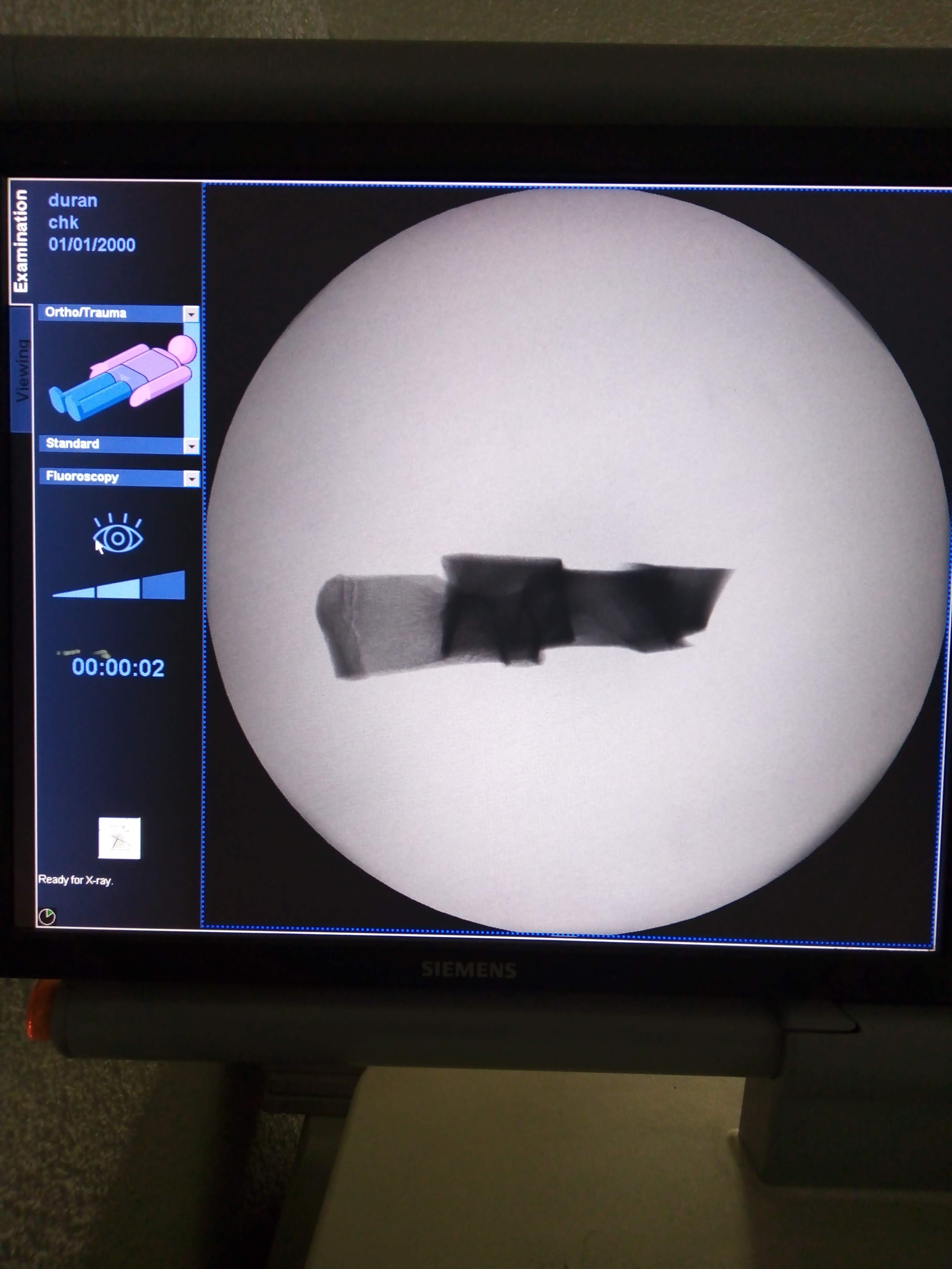
the result is an reconstructed image that is a view from the rotation axis. this means, is almost the bone was Xray'ed by the front face (not by the side).
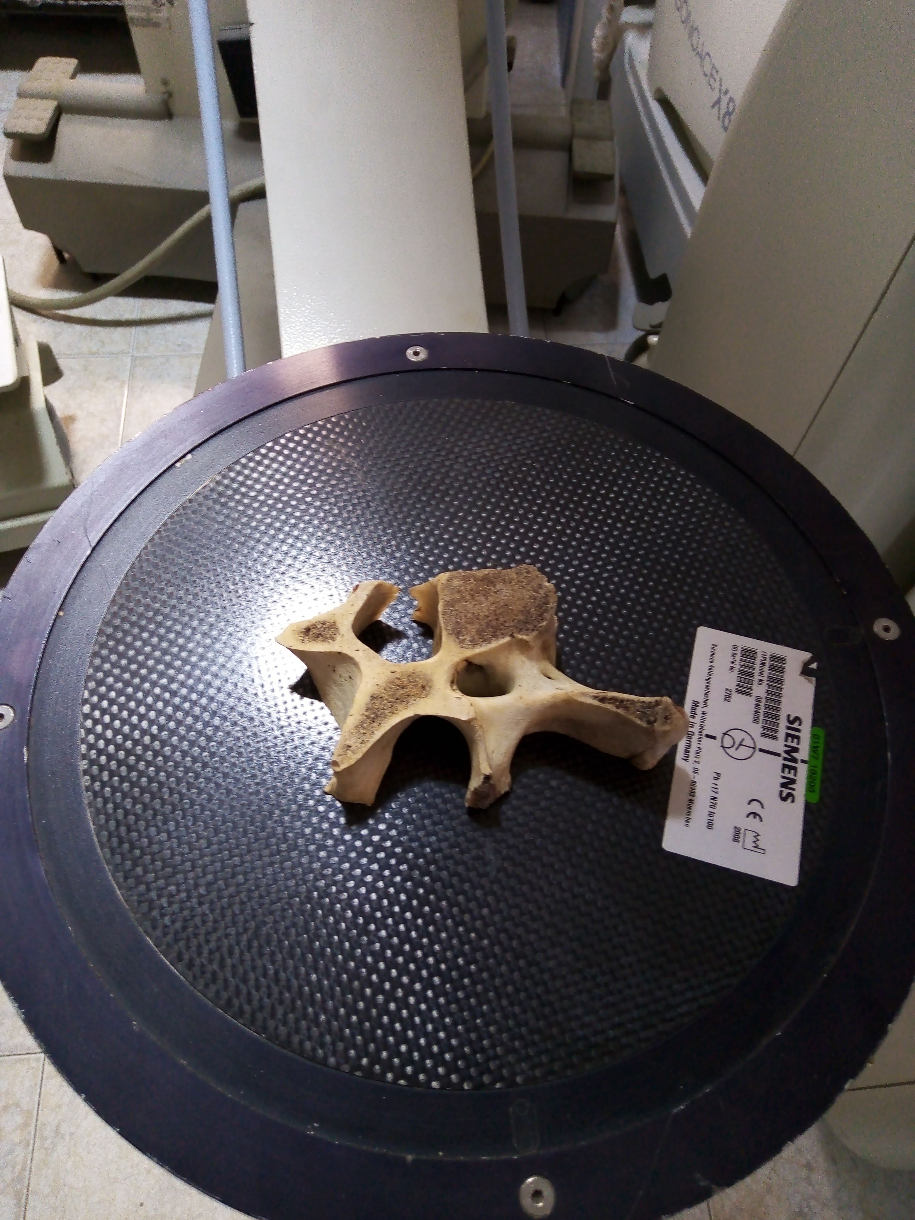
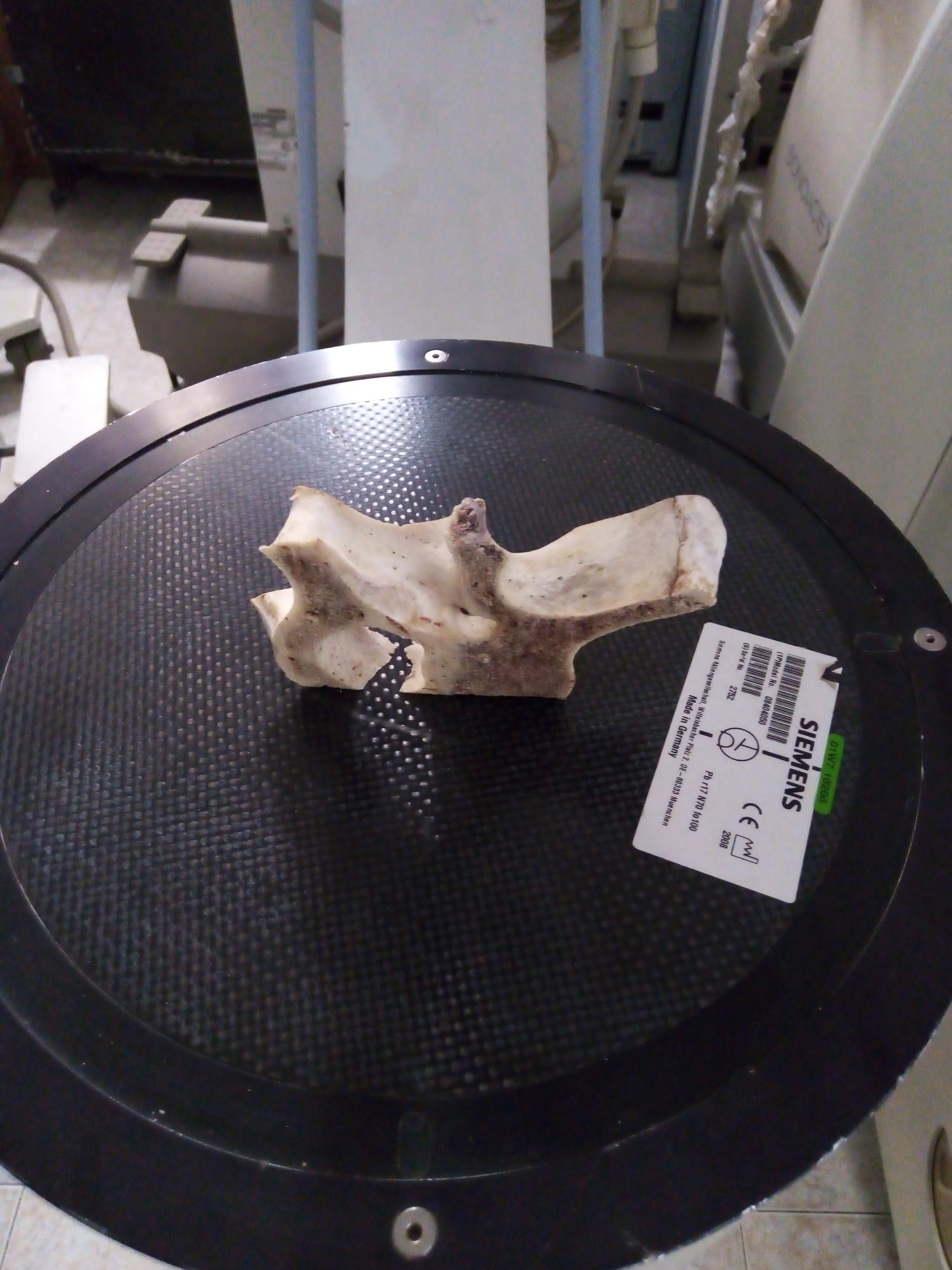
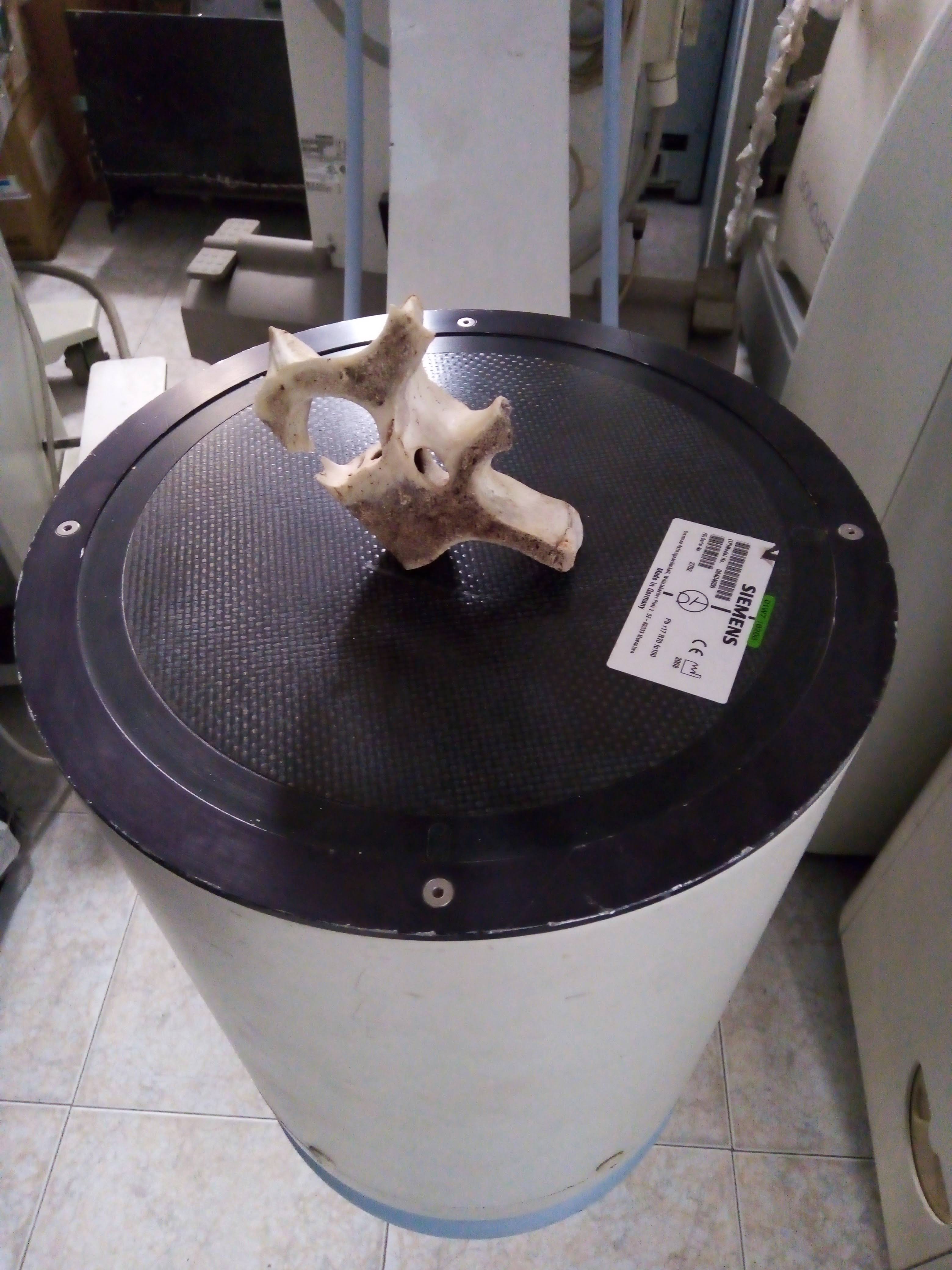
here is how image is captured in the different position. This is an arbitrary degree of rotation (CT system do this 600-1200 times in a turn), only for demo purposes.
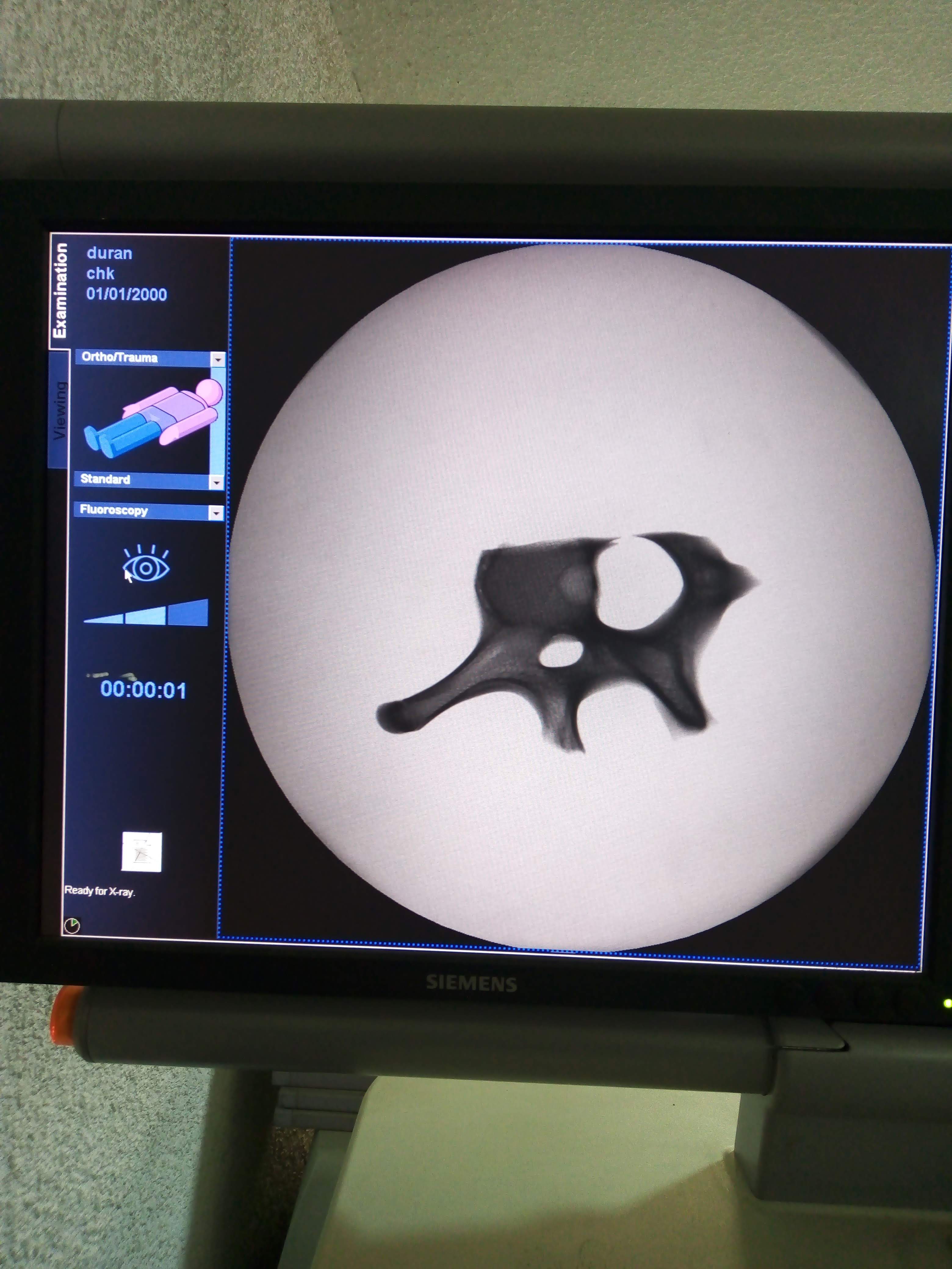
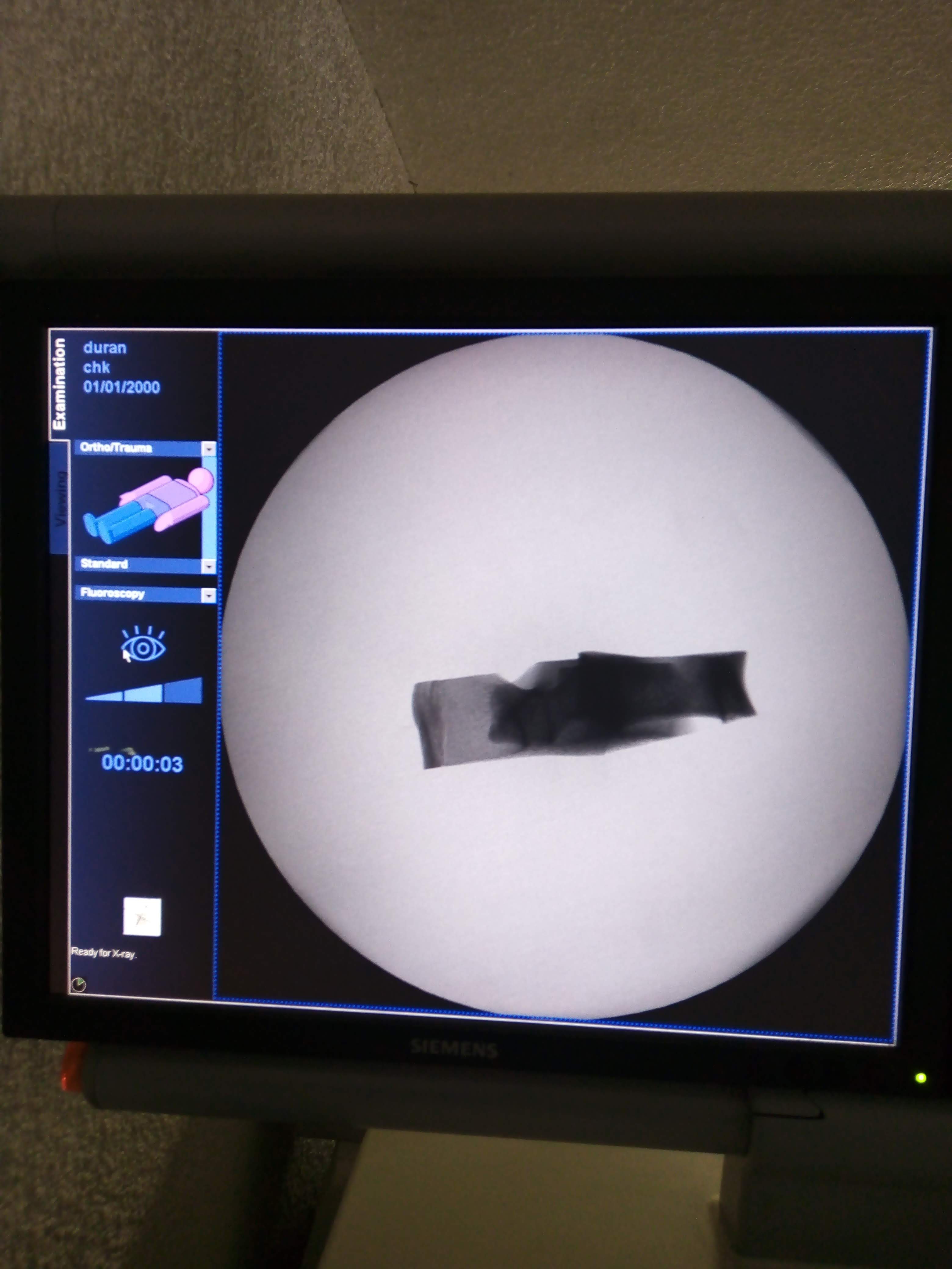
here, the Xray image produced by the cow's bone in that position.
well... to be more clear, the Xray path is from above the bone, to bottom; the dark circle where the bone is located, is the entry face of the detector. this detector is called Image Intensifier
time ago since my last entry... however, i still working on !
here, i had the chance to make some pictures in a C-arm system; C-arm is a machine with a live video that is able to deliver Xrays at one end, then capture and display the image on a monitor for reviewing or postprocessing.
here i post some to explain what CT does.
this first photo shows a cow bone i'm using as a sample/model
Well.... some time without posting here. When things needs detail, speed goes down.
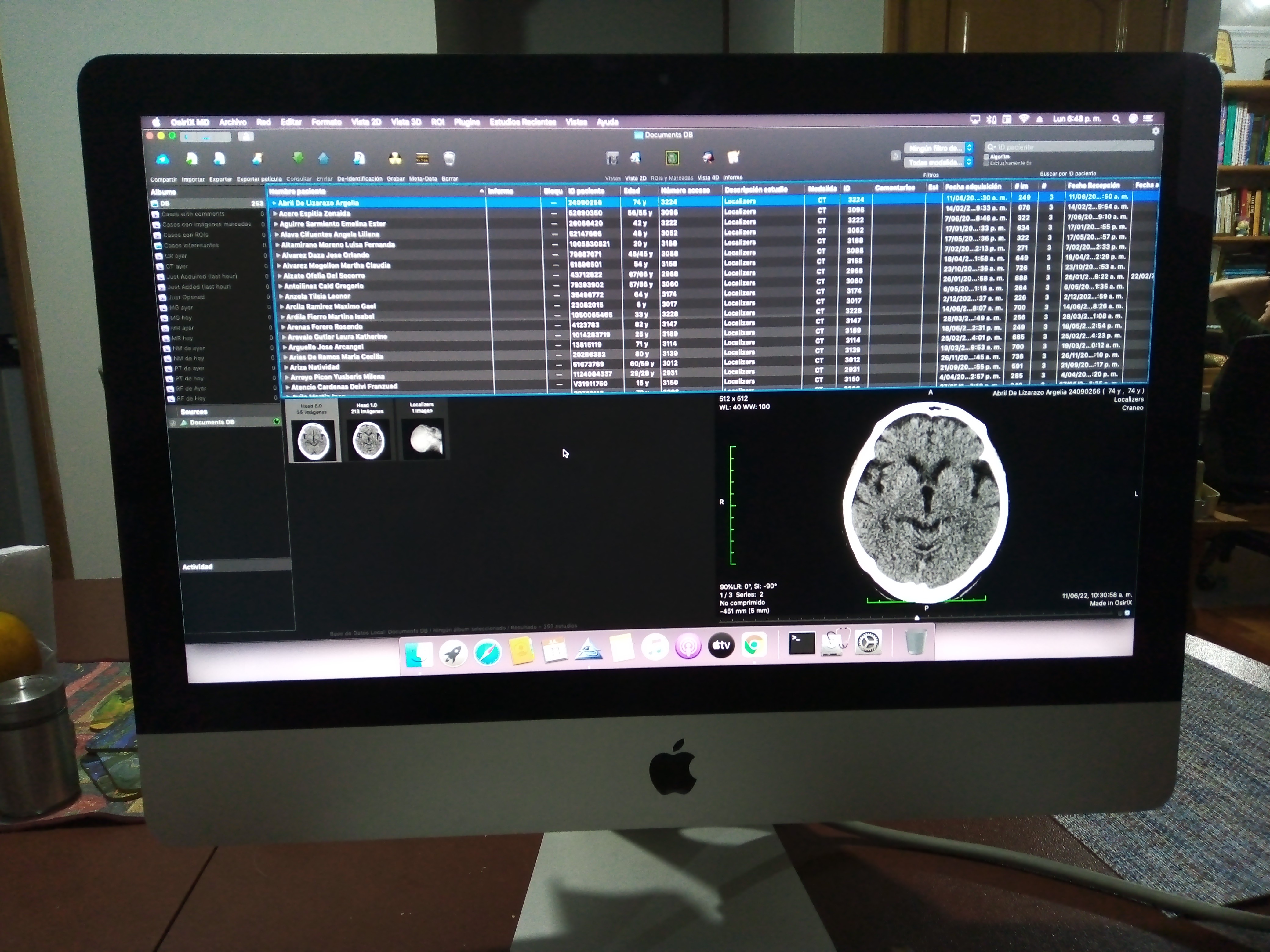
I had the chance to support a Dicom Viewer Station, implemented with an Imac PC.
Despite the commercial grade, the Retina Display has good enough specs near to Medical grade monitors, with a fraction of the cost. The picture shows the GUI from this Dicom Viewer to compare with the posted photo of the Skull; this bring a signal that is a good choice to use it.
The 3D Slicer is perfect to show the output images for the doctor´s diagnostic. However, there is a couple of things:
1) 3D slicer works with 2D images. It can use plenty of file formats, but again, 2D images.
2) CT systems does not produce images, it delivers Projections.
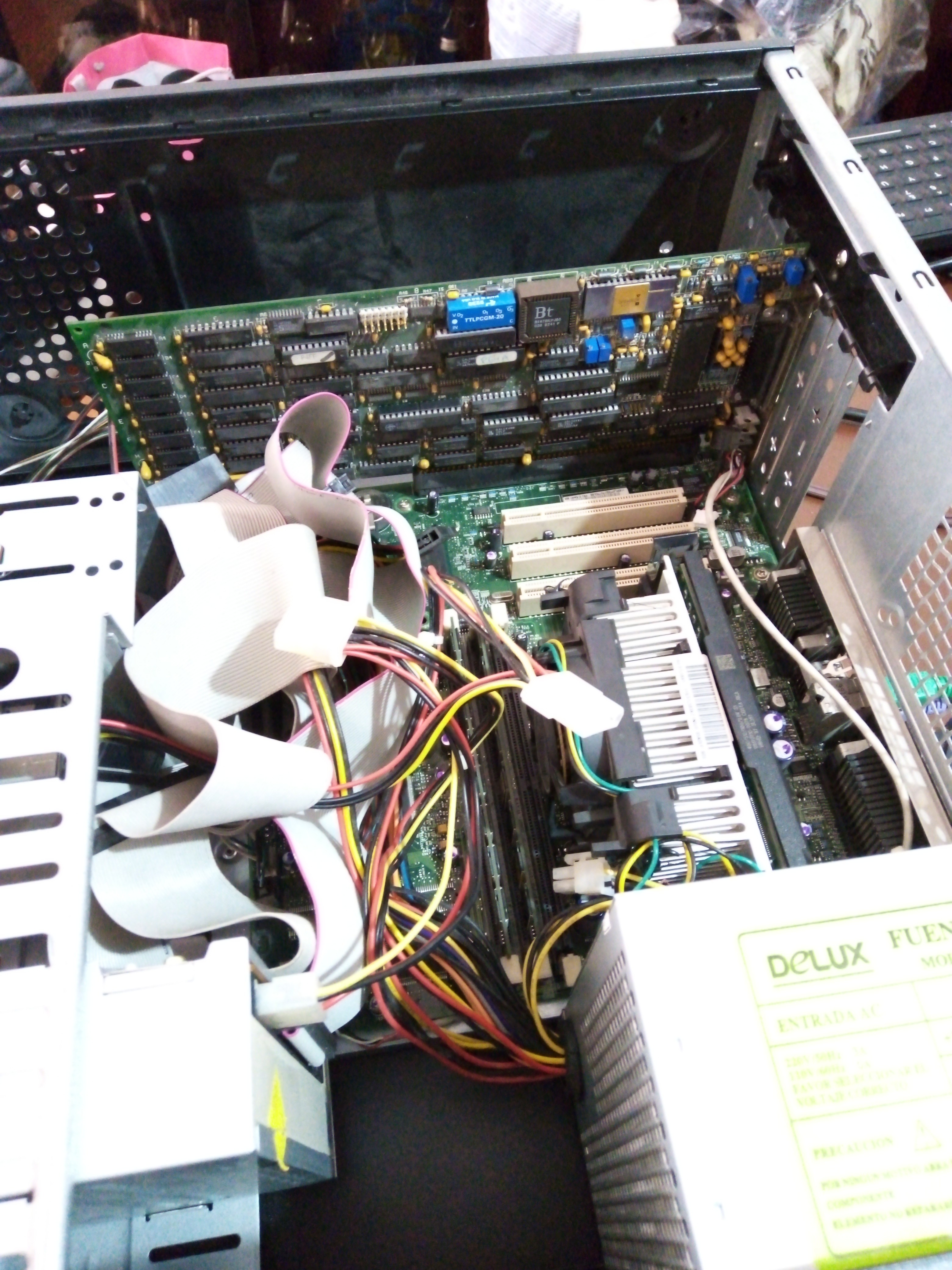
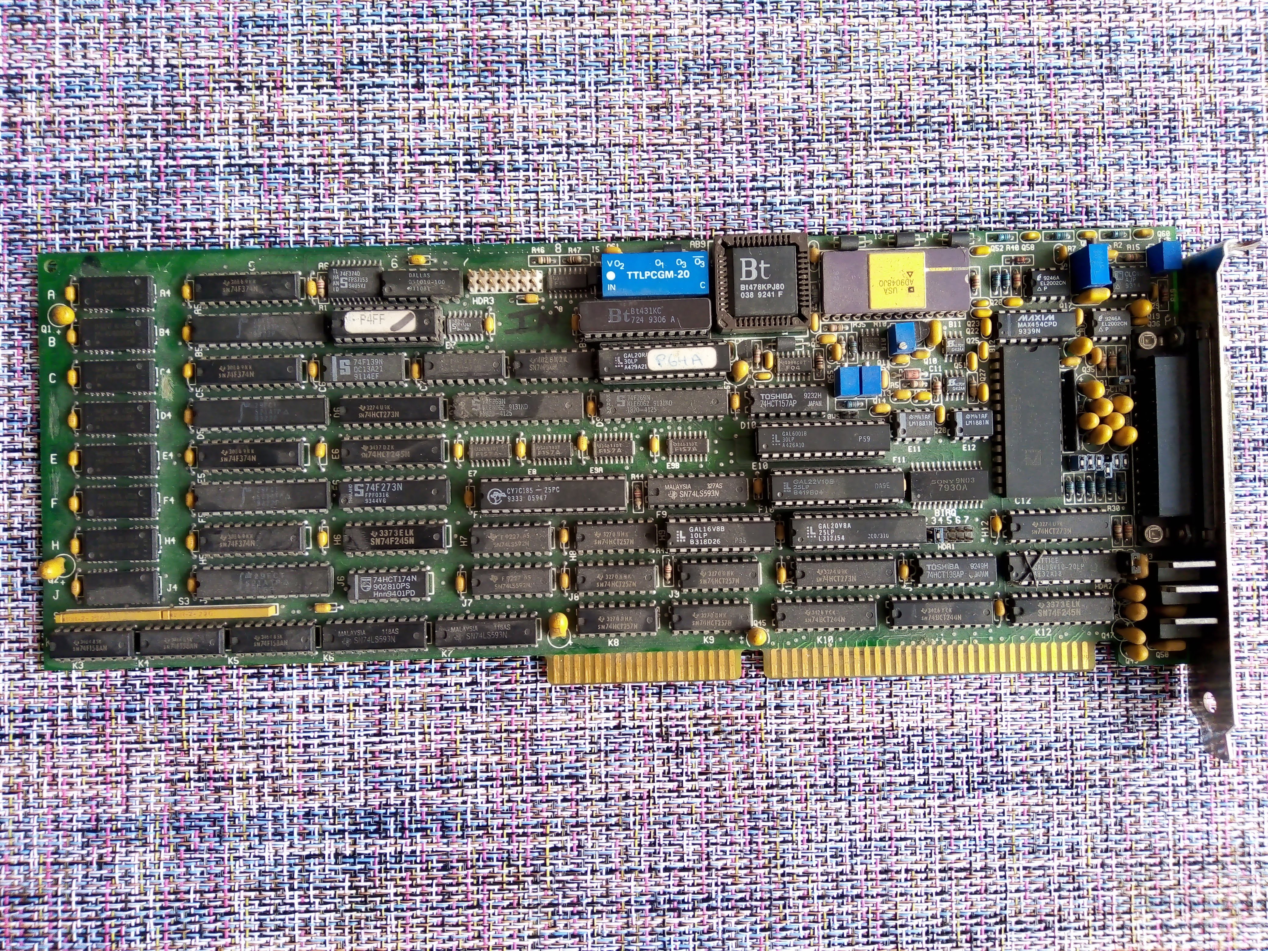
In this scenario, i need take the data collected from Gantry ( Detector), and translate to 2D images for use in 3D Slicer. To do the first part, i will Reuse this board:
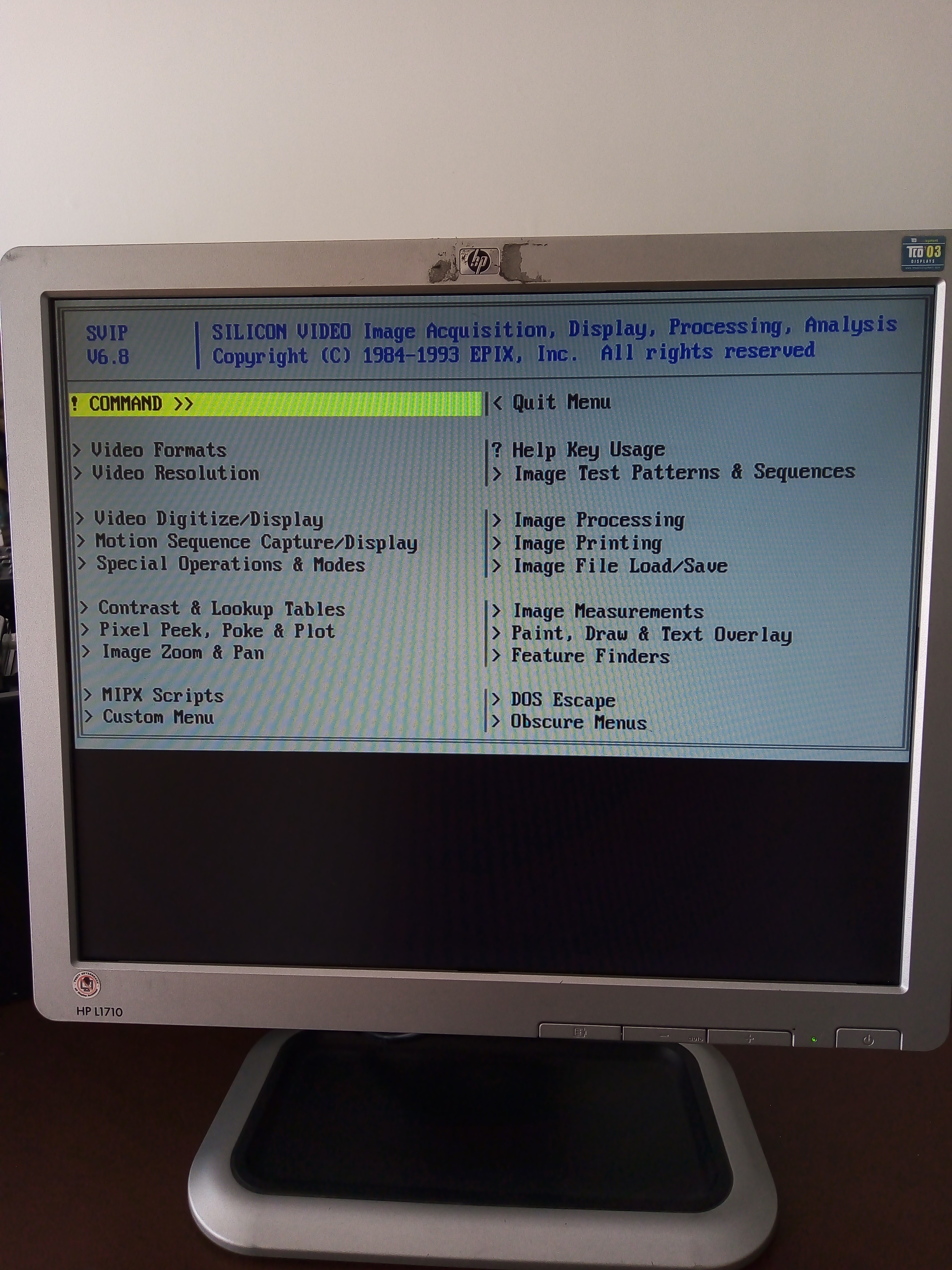
This is an old 8 bit Frame Grabber that i got, and i had used into another past projects. As you can see, this board has a ISA BUS. This is itself a challenge, because i need to put in working condition the board to capture the projections.
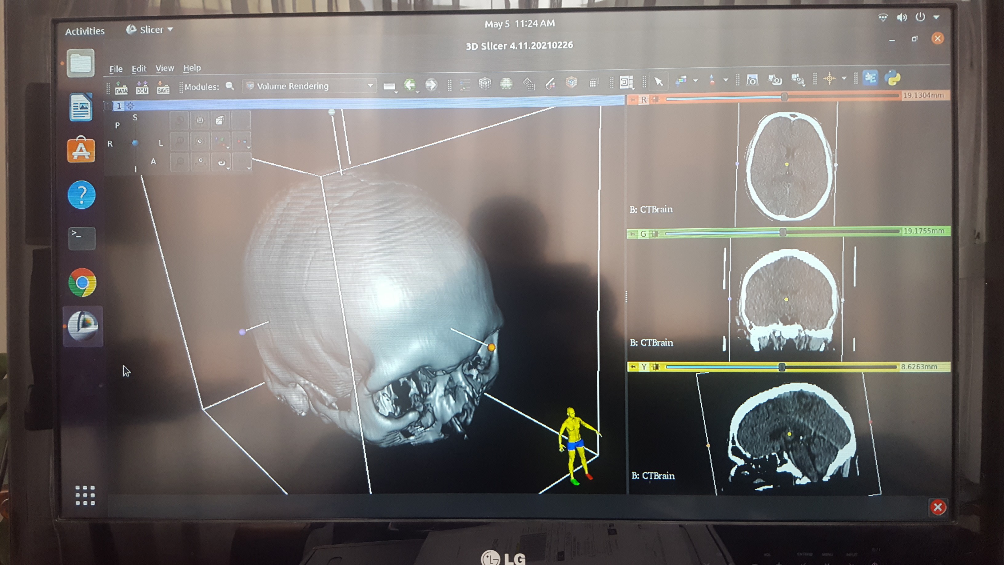
After a Tons of reading, testing and evaluating the Open Source options, i found a perfect tool for this Project (conversion of 2D images into 3D volume, then slice it arbitrary). This is exactly what a doctor does analizing the patient´s anatomy. The software with the GUI closest to market systems is 3D SLICER (www.slicer.org)
This photo shows how it works, using a DataSet available for download with the software.
keeping the Open Source soul, the system is running on UBUNTU.